Skip to content
Division of Health AIDivision of Health AI
AboutTeamResearchPublicationsJoin
Theme
Division of Health AIDivision of Health AI

Clinical AI built with the data and clinicians of Northwell Health.

Explore

  • About
  • Team
  • Research
  • Publications
  • Join

Affiliations

  • Feinstein Institutes (opens in new tab)
  • Northwell Health (opens in new tab)
  • Zucker School of MedicineHofstra Northwell

Located at

  • Institute of Health System Science
  • Institute of Bioelectronic Medicine
  • Manhasset, New York

© 2026 Division of Health AI, Northwell Health. All rights reserved.

All research

Division project · Active since 2017

Decoding the vagus nerve: preclinical recording and interpretation

Recording from the mouse vagus nerve, chronically and wirelessly, and decoding what its neurons report about inflammation and metabolism, toward bioelectronic devices that diagnose and treat from neural signals.
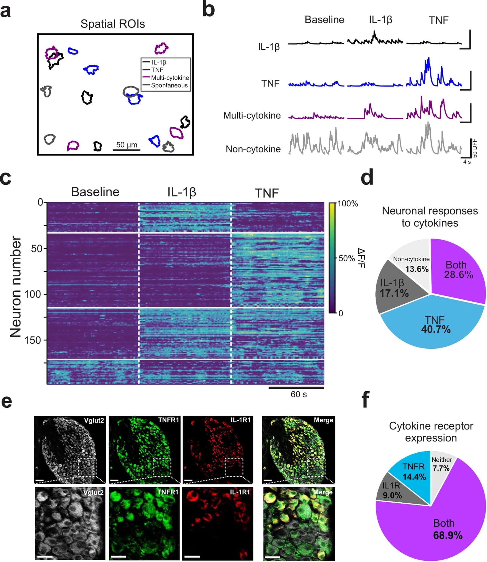
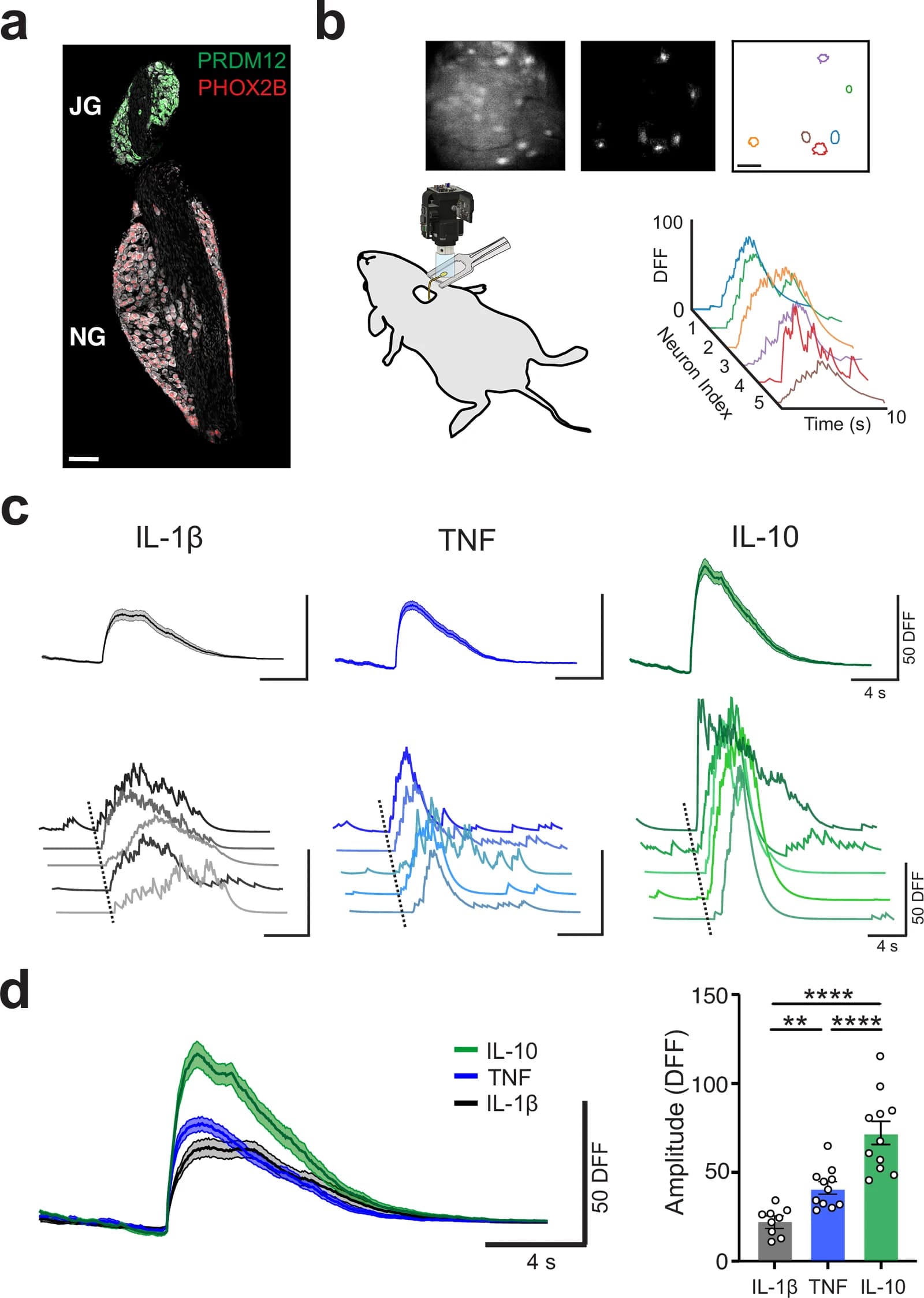
Population responses of nodose ganglia reveal subsets of cytokine-specific neurons alongside neurons that respond to several cytokines.
Population responses of nodose ganglia reveal subsets of cytokine-specific neurons alongside neurons that respond to several cytokines.Nature Communications, 2025 · CC BY 4.0 (opens in new tab)

Overview

The vagus nerve is the body's sensory channel from the organs to the brain. Its sensory neurons in the nodose ganglia respond to circulating cytokines in a cytokine-specific way, and their population activity changes in an inflamed state; the same nerve carries signals specific to hypoglycemia. The program records these signals and builds decoders that read them in real time.

Doing so in a living animal over months required new tools: a chronic implant model for the mouse vagus nerve, a fully implantable wireless bidirectional neuromodulation system, impedance-matching algorithms that remove common-mode interference, and calcium imaging of the jugular-nodose ganglia to identify distinct populations of sensory neurons.

Status
Active since 2017
Program
Division project
Publications
11, latest 2025
Team
3 members

Lines of work

3 strands

  1. 01

    Cytokine decoding

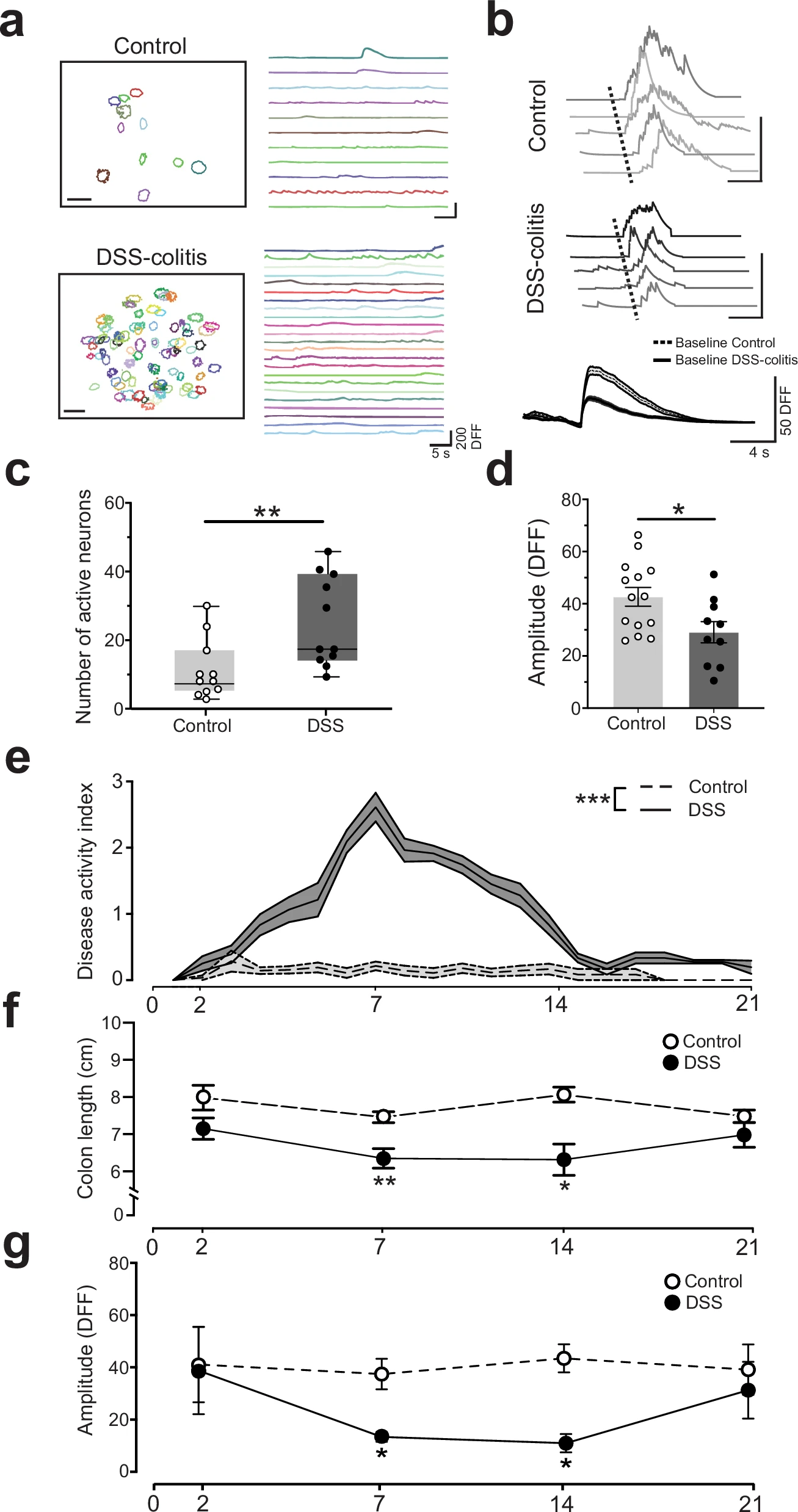
    Vagal sensory neurons represent individual cytokines; inflammation increases the number of active neurons and reduces their responses.

    Nature CommunicationsApr 2025

    Neural representation of cytokines by vagal sensory neurons

    (opens in new tab)
    Cold Spring Harbor Perspectives in MedicineJan 2019

    Recording and Decoding of Vagal Neural Signals Related to Changes in Physiological Parameters and Biomarkers of Disease

    (opens in new tab)
    Proceedings of the National Academy of SciencesMay 2018

    Decoding cytokine specific neural activity from the cervical vagus nerve

    (opens in new tab)
  2. 02

    Metabolic decoding

    Hypoglycemia-specific neural signals identified by decoding murine vagus nerve activity.

    Bioelectronic MedicineJul 2019

    Identification of hypoglycemia-specific neural signals by decoding murine vagus nerve activity

    (opens in new tab)
  3. 03

    Chronic recording and methods

    Implants, wireless systems, and signal-processing methods for months-long recording from the mouse vagus nerve.

    International Journal of Neural SystemsDec 2025

    Longitudinal characterization of compound action potentials in chronic vagus nerve recordings in mice

    (opens in new tab)
    Journal of Neural EngineeringMar 2023

    Calcium imaging and analysis of the jugular-nodose ganglia enables identification of distinct vagal sensory neuron subsets

    (opens in new tab)
    Biosensors and BioelectronicsDec 2021

    A fully implantable wireless bidirectional neuromodulation system for mice

    (opens in new tab)
    eLifeApr 2021

    Development and characterization of a chronic implant mouse model for vagus nerve stimulation

    (opens in new tab)
    Brain StimulationSep 2020

    Quantitative estimation of nerve fiber engagement by vagus nerve stimulation using physiological markers

    (opens in new tab)
    Journal of Neuroscience MethodsOct 2019

    An impedance matching algorithm for common-mode interference removal in vagus nerve recordings

    (opens in new tab)
    International Conference on Solid-State Sensors, Actuators and Microsystems (TRANSDUCERS)Jun 2017

    A new 3D self-adaptive nerve electrode for high density peripheral nerve stimulation and recording

    (opens in new tab)
Population responses of nodose ganglia reveal subsets of cytokine-specific neurons alongside neurons that respond to several cytokines.
Population responses of nodose ganglia reveal subsets of cytokine-specific neurons alongside neurons that respond to several cytokines. Nature Communications, 2025 (opens in new tab) · CC BY 4.0
Individual nodose ganglia sensory neurons respond in a cytokine-specific manner.
Individual nodose ganglia sensory neurons respond in a cytokine-specific manner. Nature Communications, 2025 (opens in new tab) · CC BY 4.0
An inflammatory state increases the number of active nodose ganglia neurons but decreases their response levels.
An inflammatory state increases the number of active nodose ganglia neurons but decreases their response levels. Nature Communications, 2025 (opens in new tab) · CC BY 4.0
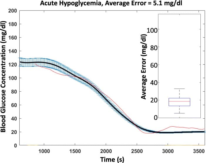
Measured blood glucose versus glucose reconstructed from vagal nerve signals during insulin-induced hypoglycemia.
Measured blood glucose versus glucose reconstructed from vagal nerve signals during insulin-induced hypoglycemia. Bioelectronic Medicine, 2019 (opens in new tab) · CC BY 4.0
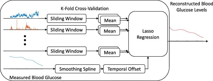
The neural decoder: sliding windows of compound action potential rates fed through lasso regression with cross-validation.
The neural decoder: sliding windows of compound action potential rates fed through lasso regression with cross-validation. Bioelectronic Medicine, 2019 (opens in new tab) · CC BY 4.0
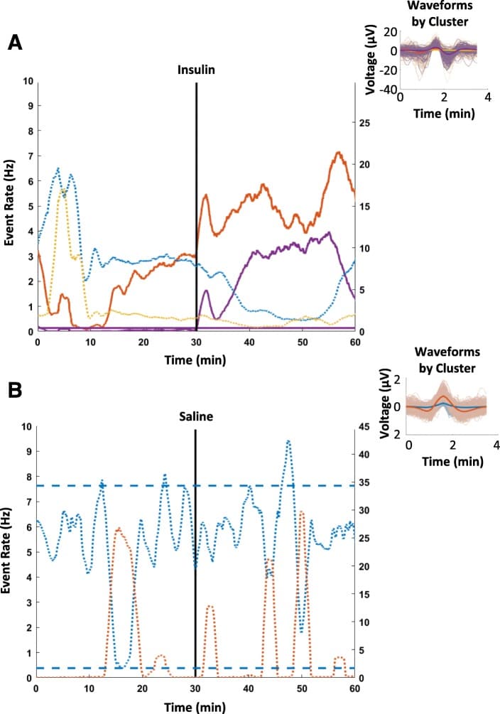
Compound action potential rates rise after insulin but not saline, showing the vagal response is specific to hypoglycemia.
Compound action potential rates rise after insulin but not saline, showing the vagal response is specific to hypoglycemia. Bioelectronic Medicine, 2019 (opens in new tab) · CC BY 4.0
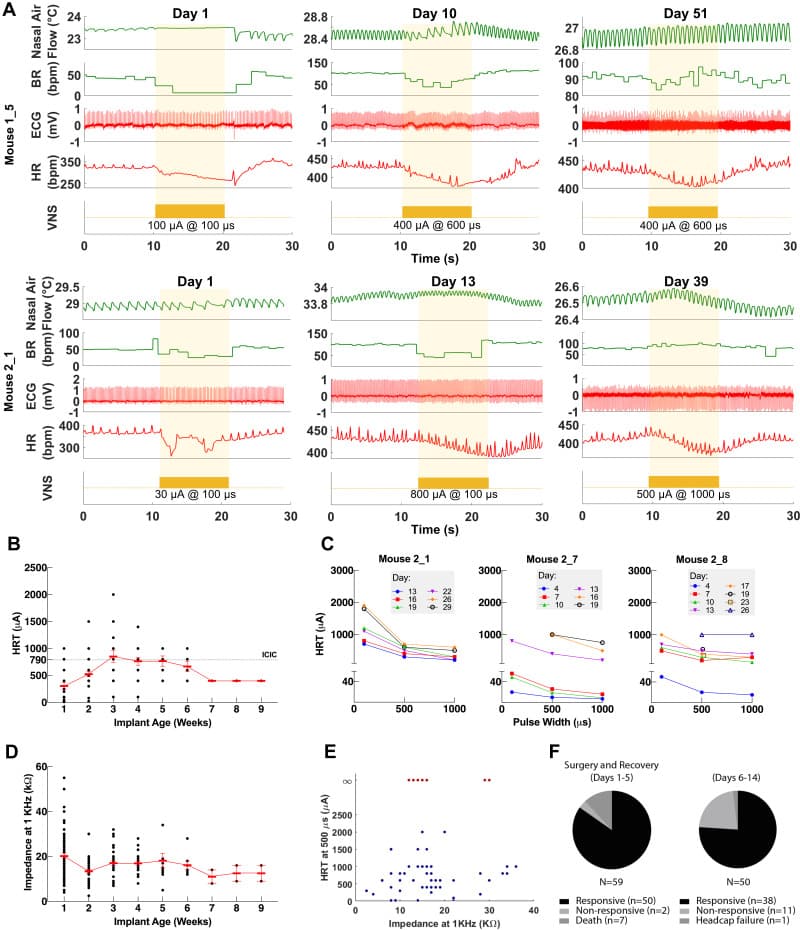
Heart rate threshold and electrical impedance stay stable for eight or more weeks after implantation.
Heart rate threshold and electrical impedance stay stable for eight or more weeks after implantation. eLife, 2021 (opens in new tab) · CC BY 4.0
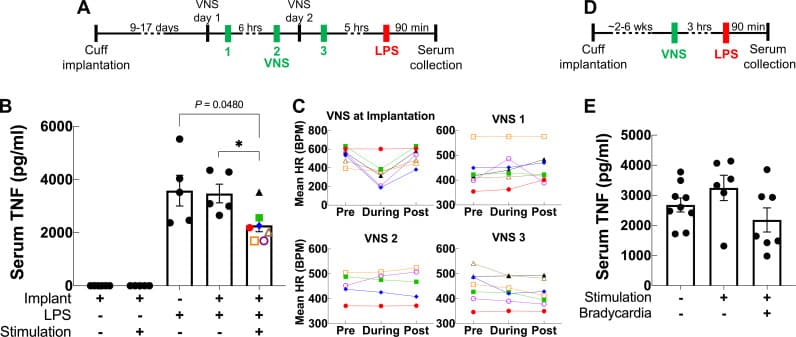
Vagus nerve stimulation suppresses serum TNF in endotoxemia versus sham in chronically implanted animals.
Vagus nerve stimulation suppresses serum TNF in endotoxemia versus sham in chronically implanted animals. eLife, 2021 (opens in new tab) · CC BY 4.0
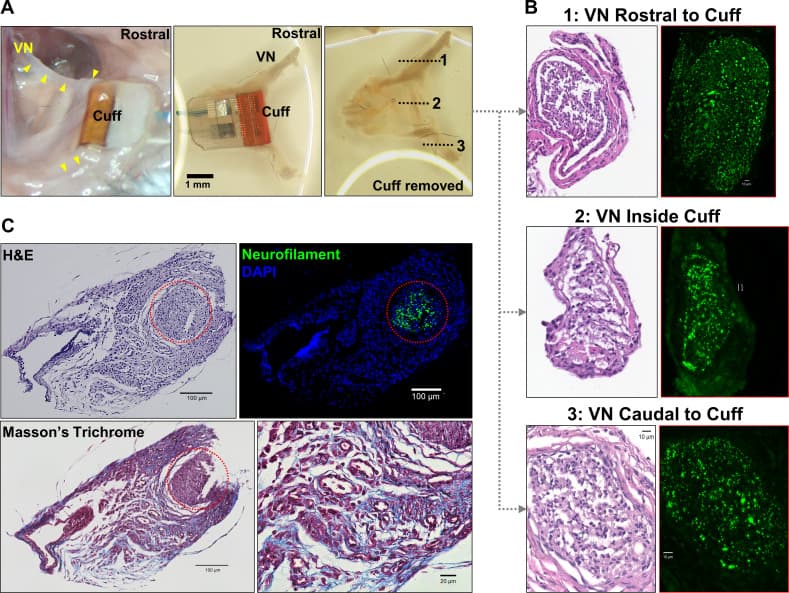
Histology of the implanted vagus nerve shows preserved fibers and intact neurofilaments at three weeks.
Histology of the implanted vagus nerve shows preserved fibers and intact neurofilaments at three weeks. eLife, 2021 (opens in new tab) · CC BY 4.0

From the papers

Published figures

Figures reproduced from the papers behind this project.

Each panel keeps its original source and license.

People

On this project

Theodoros Zanos, PhD

Professor & AVP

Shubham Debnath, PhD

Senior Research Scientist

Todd Levy, MS

Senior Biomedical Engineer

More division projects

Division project

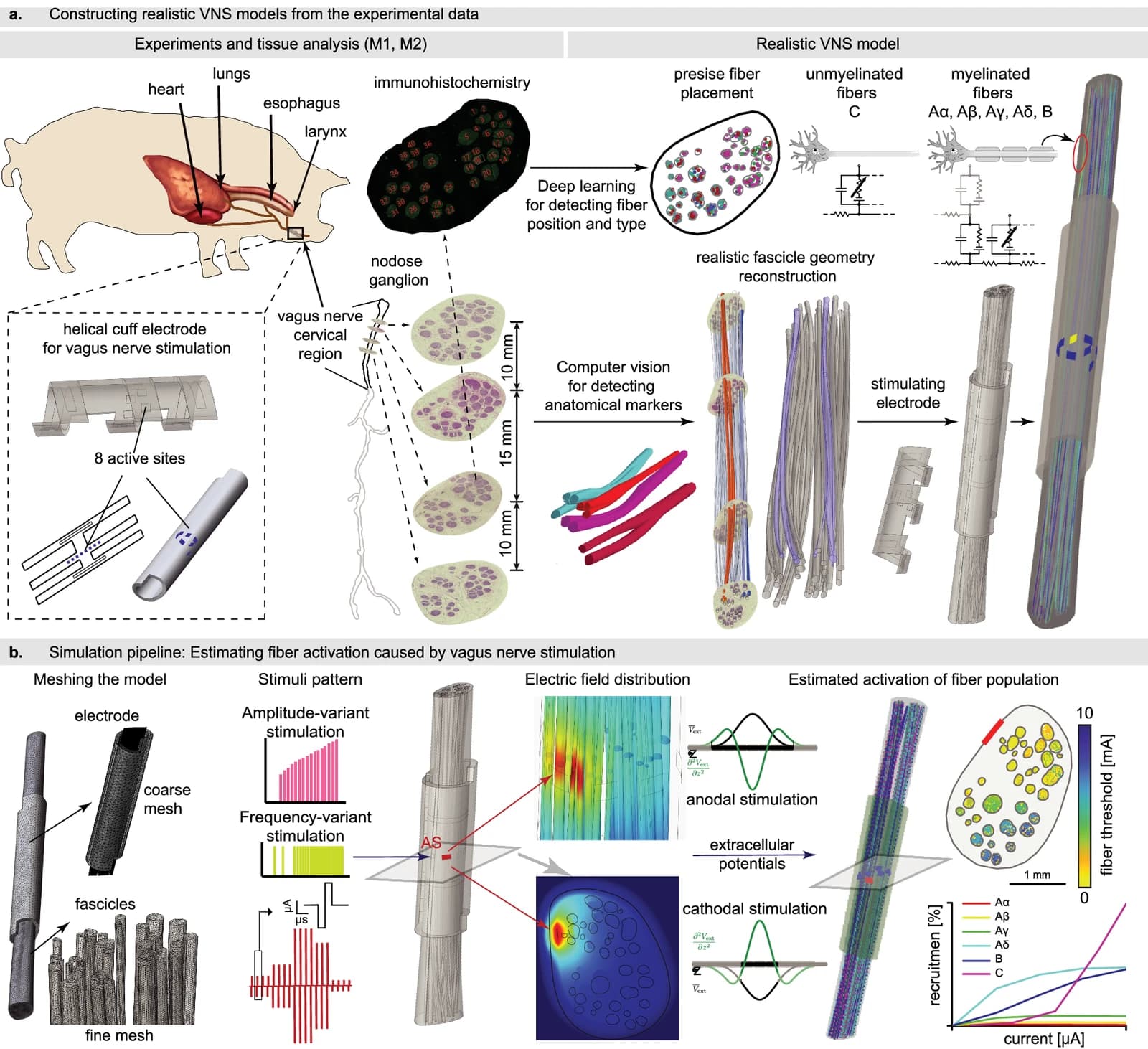
Computational models of vagus nerve stimulation

Histologically realistic models of the cervical vagus nerve that predict which fibers a stimulation pattern will activate, used to design selective, organ-specific vagus nerve stimulation.

Division project

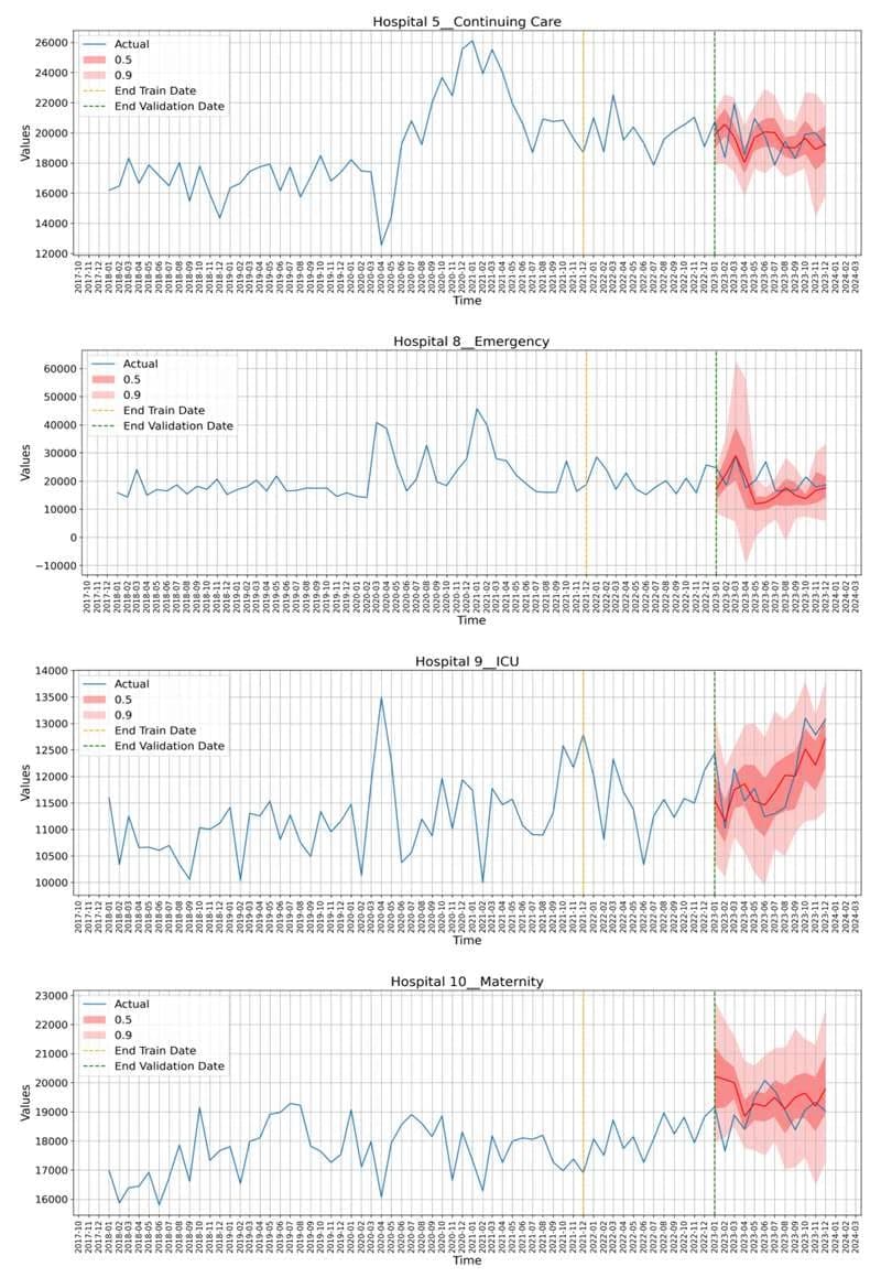
Nursing workforce forecasting

Machine learning models using DeepAR probabilistic forecasting predict nursing workforce demand across Northwell's hospital units up to 12 months ahead, supporting preemptive hiring and staffing decisions across diverse specialties.

Division project

ECG deep learning

Generative and predictive models over the electrocardiogram: reconstructing the full twelve-lead ECG from a minimal set of leads, and stratifying acute coronary syndrome risk from the admission ECG.

Division project

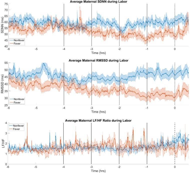
Maternal fever / neonatal sepsis prediction

Continuously monitored vital signs and heart rate variability during labor predict maternal fever 2-3 hours before clinical onset, with area under the curve of 0.748, enabling early detection of mothers at risk for neonatal early-onset sepsis.

←Previous

Computational models of vagus nerve stimulation

Next→

In-hospital deterioration prediction