Brain Stimulation
NIH-funded · Active since 2020
A comprehensive anatomical dataset of 60 human vagus nerves (30 left, 30 right), spanning millions of micro-CT images across hundreds of terabytes of data. Using 3D nnU-Net segmentation, this project builds a detailed vagus digital twin to guide the design of selective vagus nerve stimulation therapies, part of a $6.7 million NIH SPARC award in collaboration with the TNP Lab.
Overview
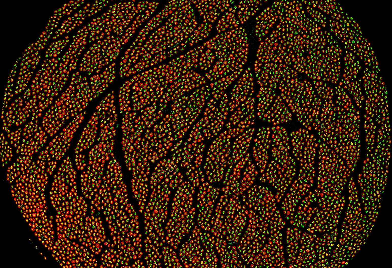
The vagus nerve carries the signals that bioelectronic medicine aims to read and write, and until REVA no complete map of its fibers existed. Reconstructing Vagal Anatomy (REVA) imaged 60 human vagus nerves, 30 left and 30 right, with micro-CT at fiber resolution, producing millions of images and hundreds of terabytes of data, and segmented them with 3D nnU-Net into a digital twin of the nerve from the neck to the abdomen.
The maps were released publicly through the SPARC data portal in July 2026. They show how organ-specific fascicles, such as the bronchopulmonary and recurrent laryngeal fibers, are organized along the nerve, which is the anatomical basis for stimulation that reaches one organ and spares another.
REVA is a $6.7M NIH SPARC award to the Feinstein Institutes for Medical Research, with the division responsible for the segmentation and analysis pipeline, in collaboration with the TNP Lab.
- Status
- Active since 2020
- Program
- NIH-funded
- Funding
- NIH Common Fund, Stimulating Peripheral Activity to Relieve Conditions (SPARC)
- Publications
- 2, latest 2023
- Team
- 3 members
Awards
Reconstructing Vagal Anatomy (REVA): a comprehensive anatomical map of the human vagus nerve
NIH Common Fund, Stimulating Peripheral Activity to Relieve Conditions (SPARC) · $6.7M · Multi-year award
Feinstein Institutes for Medical Research
NIH SPARC program (opens in new tab)
Publications
Papers
Annual International Conference of the IEEE Engineering in Medicine and Biology Society (EMBC)
People